The 2019 AAMD Contouring Session - A Primer
Goals
This session will be both practical and immediately applicable to your work. The goals are to assess and improve contouring skills by combining hands-on contouring exercises, objective analysis of accuracy and variation, and physician lectures.
In this session, we will focus on three important anatomical structures of the male pelvis: prostate, seminal vesicles, and penile bulb.
Learning Strategy
The learning strategy combines complementary components:
- Manual contouring by each participant of each of the anatomical structures for two different patient datasets (pre-meeting exercises are open to everyone; live session exercises are available to meeting attendees who bring WiFi-enabled laptops and mouse)
- Individual comparison and analysis of your contours vs. the consensus physician lecturer’s contours via private, interactive, online tools
- Analysis of population statistics and inter-observer variation using 3D consensus visualization as well as intuitive charts and basic statistics
- Physician team lectures on each structure focusing on methods, protocols, tips and techniques, as well as commentary on how they created consensus contours and implications of variation
- Question and Answer with the physicians, time permitting
Methods
Pre-Meeting Contouring Exercises
One patient imageset and contouring exercises for each structure will be open to everyone, worldwide. These will be released as structures in the ProKnow Contouring Accuracy Library and configured to be available to anyone with a ProKnow Quality Systems account (setting up an account is free and takes just a moment).
These structures will be released by June 1, 2019, leaving plenty of time to contour each of the three structures prior to the AAMD meeting.
Live Session Contouring Exercises & Lectures
The session is part of the General Session agenda on Sunday, June 16. During the session, three new contouring exercises – for the same anatomical structures but for a different patient dataset – will be released during the live session at the AAMD meeting. These exercises will be available to attendees of the meeting who bring a WiFi-enabled laptop and mouse or other pointing device (Note: Mac and Android tablets will not work).
For each structure, the workshop will follow this process:
- Analysis of pre-meeting contouring exercise results, focusing on inter-observer variation and consensus views
- Lectures and commentary by the physician team for the structure, including published methods as well as other tips and techniques
- Live contouring of the same structure on the new dataset
- Real-time analysis of results of the live contouring exercise
Physician Lecturers

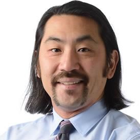
Aaron Kusano, MD is a radiation oncologist at Anchorage and Valley Radiation Therapy Centers in Alaska. During his residency at the University of Washington, he realized that good radiation plans relied as much on objectives and contours as they did technology. Dr. Kusano continues a longstanding interest in quality and safety in radiation oncology in his busy community practice as the Medical Director, physician lead for RO-ILS and head of gynecologic brachytherapy program.

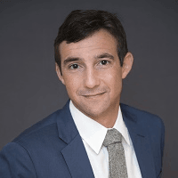
Matthew Ladra, MD is an assistant professor of Radiation Oncology at the Johns Hopkins School of Medicine in collaboration with Children’s National Medical Center in Washington DC. He completed his radiation oncology residency at the University of Washington Medical Center and a 2-year pediatric proton fellowship at Massachusetts General Hospital. Prior to joining Johns Hopkins Dr. Ladra served as the pediatric director of radiation oncology at the Provision Proton Therapy Center in Knoxville TN.