The case for MR-to-MR deformable image registration with Elekta Unity MR-Linac
Study evaluates accuracy of Elekta’s deformable registration algorithm for both CT-MR and MR-MR registrations
The superior soft-tissue visualization of MR-guided radiotherapy offers the potential to greatly increase the accuracy of each patient’s treatment fraction. However, this requires the targets and organs-at-risk (OARs) to be defined on the daily MRI scan. This task of re-delineating and validating targets and organs-at-risk (OARs) can be time-consuming and increases the risk of patient motion during the treatment session.
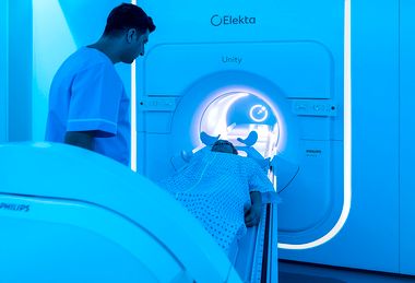
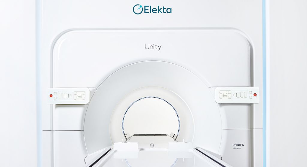
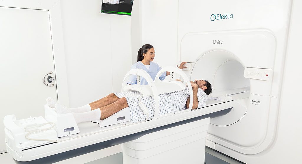
To address this issue, Elekta Unity users employ an automatic deformable image registration (DIR) algorithm in the MR-Linac’s planning system to speed deformation of the planning images and structure propagation. To determine which registrations are more accurate – CT-to-MR or MR-to-MR – Odense University Hospital researchers compared them to benchmark (i.e., “ground truth”) manual delineations.
The study involved 12 high-risk prostate cancer patients, for whom target structures and OARs were delineated on both planning MR and CT scans and propagated using DIR to three T2-weighted MR scans acquired during the treatment course1. The investigators assessed the structures generated against manual delineations on the repeated scans employing intra-observer variation on the planning MR as the benchmark.
The criteria used to evaluate the accuracy of DIR were:
- Dice similarity coefficient (DSC): The ratio of overlap between the manually delineated structure and the corresponding deformable propagated structure. This is most relevant for smaller structures.
- Mean surface distance (MSD): The average distance between the manual and deformed structure in absolute measures, which is especially relevant for larger structures.
- Hausdorff distance (HD): Delivers the greatest distance between a given pair of structures to show a worst-case scenario, thereby making it sensitive to outliers in the data.
For each patient, the average value over all scans of the DSC, MSD and HD was calculated for each structure investigated for both MR-to-MR and CT-to-MR registrations and compared to the intra-observer variation.
The results demonstrated that all of the MR-to-MR propagated structures yielded higher median DSC than CT-to-MR propagations when compared to the benchmark manual delineations, suggesting that MR-to-MR DIR is more accurate. This finding was statistically significant for the prostate, seminal vesicles, rectum, femoral heads and penile bulb.
The MSD values showed better agreement with the manual delineations for all deformed structures based on MR relative to CT.
In addition, overall, MR-to-MR deformed structures largely showed DSC and MSD values in the same range as the intra-observer variations. MR-to-MR DIR yielded smaller HD values for all eight investigated structures than CT-to-MR. DSC and MSD also showed a statistically significant difference between CT-to-MR propagated contours and the intra-observer variation for all organs.
“MR-to-MR was statistically similar to the intra-observer variation in the majority of cases.”
Importantly, MR-to-MR was statistically similar to the intra-observer variation in the majority of cases.
“These results were so good that they are comparable to the variation among expert users,” says Kevin Brown, Distinguished Scientist at Elekta. “You can’t hope for a much better outcome.”
Brown adds that this study showed that Elekta’s DIR works better for smaller changes than larger changes in targets and OARs.
“Elekta Unity users are often building this factor into their workflow,” he notes. “The system enables the user to choose any of the previous treatment sessions to be the reference images. Therefore, users are simply choosing previous MR scans that are similar to the current scan in order to improve the results.”
Although the researchers conclude that MR-to-MR propagated structures require fewer corrections and are thus preferred for clinical use over CT-to-MR propagated structures, Elekta Unity users have options regarding which imaging modality to use for planning.
“Elekta Unity supports either MR-to-MR or CT-to-MR workflows,” Brown says. “Unity users that have only CT simulation at their disposal sometimes use the first treatment session to acquire an MR that they can use in subsequent sessions for this registration.
“Throughout the coming years, we anticipate that the availability of high-quality data from ongoing patient treatments will drive AI image segmentation that will be fast and accurate, significantly reducing treatment time,” he adds. “However, this study demonstrates that the current structure propagation tools integrated in Elekta Unity are useful and practical.”
Learn more about Elekta Unity.
- Christiansen, RL, Dysager, L, Bertelsen, AS et al. Accuracy of automatic deformable structure propagation for high-field MRI guided prostate radiotherapy. Radiat Oncol 15, 32 (2020). https://doi.org/10.1186/s13014-020-1482-y
