Imaging improvements expand clinical scope at Australia hospital

RTTs and physicists report better soft tissue visualization, benefiting registration and alignment for several treatment sites
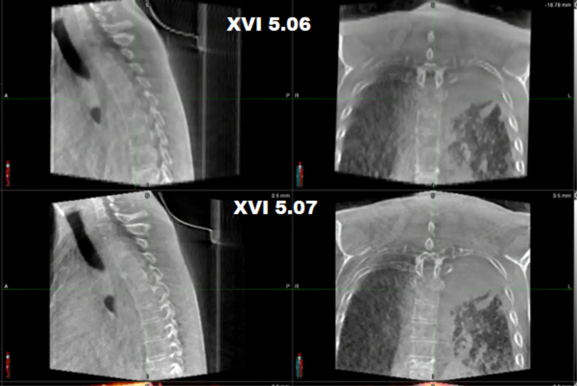
Elekta’s latest upgrade to the XVI (v. 5.0.7.) cone beam CT (CBCT) imaging workflow in its Versa HD™ system is having a noticeable impact, according to RTTs at Flinders Private Hospital (GenesisCare, Bedford Park). They report that they can more efficiently register targets and organs-at-risk and are more confident in treatment verification. They also note that the imaging improvements could potentially expand their center’s clinical scope. The upgrade was included in the Versa HD system the clinic began using in March 2023, the center’s second linac.
I’ve been on this Versa HD since we went live and instantly it was very obvious that the soft tissue visualization was a substantial improvement.”
“I’ve been on this Versa HD since we went live and instantly it was very obvious that the soft tissue visualization was a substantial improvement,” says Lauren Knight, RTT, Radiation Therapist Unit Leader. “We do a soft tissue match with our prostate patients and it’s so much clearer than the images on the other linac. The edges are more sharply defined, I can easily delineate the most posterior part of the bladder and where the prostate starts, and the anterior rectal wall is nice and clear.”

Knight has also observed sharper definition of the skin edge in breast patients, for whom the use of half gantry arcs to avoid clearance issues would sometimes cause artifacts on the skin surface when using the existing linac.
“While we still check bony alignment – the chest wall, lining up the ribs correctly, the head of the humerus, the shoulders – we still want to ensure we’re covering at the skin edge and that the breast tissue has fallen in the correct area where it should be lining up,” she explains. “This has been a noticeable improvement with the CBCT upgrade, especially for patients with pendulous breasts.”
Artifact reduction - through the ghost calibration and correction - is one of the upgrade’s main benefits, is not limited to soft tissues, Knight observes, but also to bone.
“Many spine patients, for example, might have metal pins through a bone, which can cause image saturation in certain regions, leading to streaking line artifacts through the image, which in turn can change the contrast, making everything show up a bit bright and confuse where the edge of the bone sits,” she says. “You can spend a lot of time modifying windows and levels to eliminate that to allow you to see what you want to match. With the XVI upgrade, there has been a notable improvement in bone visualization – the edges are a lot clearer and cleaner.”

Patients with bilateral hip replacements can benefit from artifact reduction where the implanted prosthesis can interfere with proper visualization of pelvic structures such as the prostate.
“With the upgraded system, there are appreciable image quality improvements in these patients,” Knight says. “I recall scanning one gentleman recently who had bilateral hip replacements and I could observe a reduction in gross shadowing and streaking between and around the prosthesis. The reduction in overall artifact definitely gives the treatment team greater confidence when registering our patients.”
Knight has found that the enhanced image quality also has enabled her to use a larger field-of-view in, for example, bariatric/larger patients or patients for whom they’re treating a large region, such as a bilateral chest, bilateral shoulders, or other cases in which there is a wide anatomical separation.
“It gives me greater confidence in applying larger fields-of-view and filters to patients,” she observes. “In the past we used them with caution, knowing that although a larger FOV might give you a wider perspective, it also shortens the scan.”
Equipped with better soft tissue visualization, she is looking forward to the center exploring the application of SRS for cranial targets.
“This would offer an alternative imaging pathway to our current process that employs ExacTrac® kV imaging,” Knight remarks. “But that technology just gives us 2D stereoscopic imaging for those cranial cases. Having an improved image from the cone beam would help us with the required certainty for treating very small cranial tumors while using C-RAD SGRT for tracking non-coplanar deliveries.”
High-dose SBRT for targets in the liver, pancreas, kidney and adrenal glands might also be on the horizon at her clinic, Knight adds, facilitated not only by the enhanced image quality, but also the second linac’s HexaPOD™ evo RT robotic patient positioning system.
“I know there will be benefits for SBRT of these organs,” she says. “Having previously treated these patients at a previous clinic in which I worked I am excited by the potential to perform image matches more accurately. The ability to eliminate the need to insert fiducial markers for liver patients, especially, is promising.
“The enhanced soft tissue image quality will help us, as tumors in those organs are very hard to delineate. We could start taking some of those patients at this site.”
“In the past, we had to send those patients to another site that has a suitably equipped Versa HD,” she adds. “The enhanced soft tissue image quality will help us, as tumors in those organs are very hard to delineate. We could start taking some of those patients at this site.”
A physics perspective on the latest CBCT release
The physics group, which commissioned the Versa HD and new CBCT release at Knight’s center, compared the upgrade with the previous CBCT release using Elekta’s acceptance image protocols.

“The Catphan® phantom image tests were improved across uniformity and low- and high-contrast visibility tests,” says Luis Muñoz, Senior Medical Physics Specialist. “In terms of clinical use, for patients, the soft tissue contrast and bone definition has observably improved. This can be demonstrably seen for our seedless prostates when compared to the previous CBCT release. Reconstructing between Partial2 and Bilinear shows an improvement in the image quality for our pelvis patients.”
“While for small fields-of-view there has been a nominal impact on image quality, the team has reported that for medium and large FOVs that is a noticeable image quality improvement.”
GenesisCare Head of Physics, Peter McLoone says: “While for small fields-of-view there has been a nominal impact on image quality, the team has reported that for medium and large FOVs that is a noticeable image quality improvement.”

“Having improved medium and large FOV images that can be assessed against CT in the future will be key for adaptive workflows as they’re released in the Elekta ONE treatment planning system,” Muñoz adds. “This exciting advancement marks a new era for radiotherapy on Elekta linacs, promising improved patient outcomes.”
McLoone says that GenesisCare is investigating how a commercial machine learning algorithm performs on their enhanced image quality scans and how the improvements better support the team in identifying patients who required replanning due to anatomical changes.
“RTTs need clear images so they have confidence to not only set the patient up correctly on the day of treatment, but also to request a review from the planning team when they are concerned about anatomical changes,” he says.
Learn more about advanced imaging with Versa HD.
LARVHD230905


