Functional imaging on MR-Linac provides insight on brain tumors
Elekta-sponsored webinar reviews Sunnybrook Health Sciences Centre’s experience using diffusion-weighted imaging
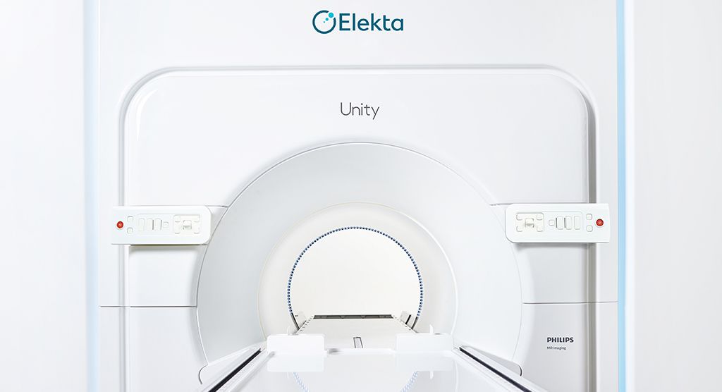
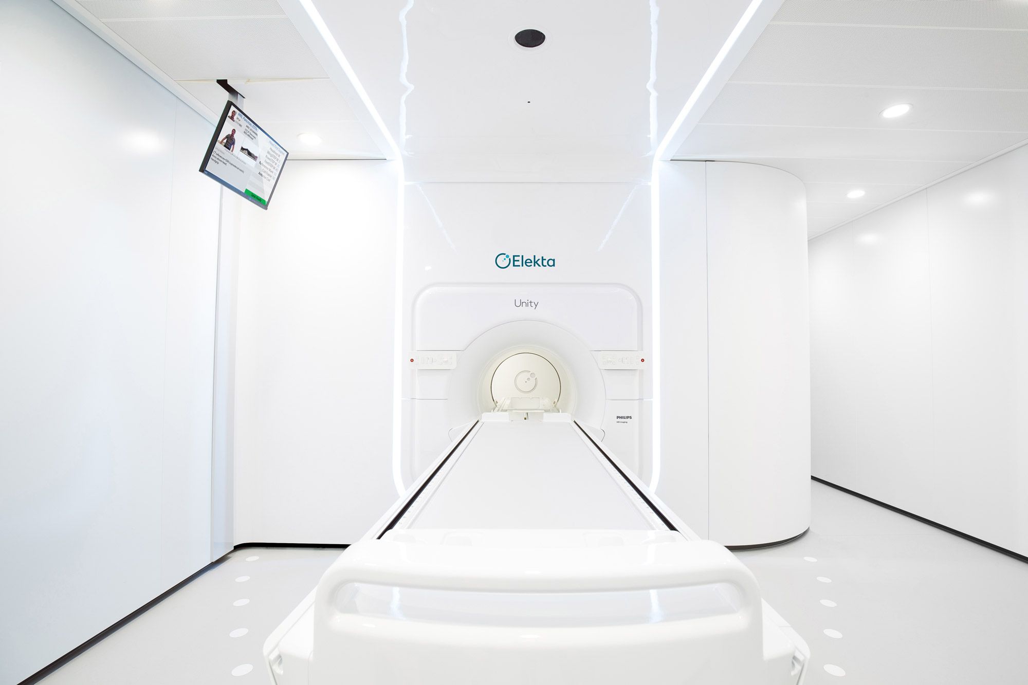
Scientists at the University of Toronto and Sunnybrook Health Sciences Centre reviewed their center’s first clinical use of the Elekta Unity MR-Linac. Chia-Lin (Eric) Tseng, MD, and Angus Lau, PhD, explained how they harnessed functional imaging MRI sequences, including diffusion-weighted imaging (DWI) to evaluate patients with CNS tumors, the majority being glioblastoma multiforme (GBM). View the full webinar.
DWI is an MRI sequence that quantifies the diffusion of water molecules to generate contrast in MRI images. DWI can be used to assess changes in tumor size and functional characteristics associated with radiotherapy or other treatments.
A founding member of the Elekta MR-Linac Consortium, Sunnybrook has used its Elekta Unity system to treat over 60 patients, 42 percent of whom have GBM, an aggressive brain tumor. The center began a prospective imaging study of GBM patients undergoing chemoradiotherapy before the Elekta Unity installation in 2017.
“The purpose of this study on the MRI simulator was to gain a prospective view of how things were changing, dynamically, in the target and organs-at risk, as patients’ go through their 30 treatment fractions,” says Sunnybrook radiation oncologist Chia-Lin (Eric) Tseng. “With Elekta Unity, some of these changes may be appreciated which we would not otherwise have been able to detect if the patient had been treated on a conventional linac.”
For GBM patients, the center uses an Adapt to Position (ATP) workflow, which registers the CT-SIM to each MRI fraction. The whole process takes about 30 minutes, but can be as low as 26 minutes without the addition of research sequences, such as magnetization transfer (MT) and chemical exchange saturation transfer (CEST), Dr. Tseng notes.
“This approach is faster than Adapt to Shape [ATS], because no recontouring by a radiation oncologist is required and no additional tissue segmentation is done on the Elekta Unity MRI, as the CT SIM already has the electron density information,” he says.
Dr. Tseng presented images of several clinical cases in which significant changes in tumor volume were observed on the MR simulator and Elekta Unity DWI datasets throughout the treatment course.
Dr. Lau, imaging scientist at the Sunnybrook Research Institute and Odette Cancer Centre, followed with a discussion of Sunnybrook’s daily CNS multi-parametric imaging protocol for the Elekta Unity; protocol optimization; geometric distortion correction; validation by comparison to a MRI simulator; initial results with daily apparent diffusion coefficient (ADC) measurements; and comparison between pre-beam, beam-on and post-beam scans.
“In protocol optimization, the intent is to maximize signal-to-noise and contrast-to-noise ratios,” Dr. Lau says. “We spent a lot of time thinking about how to minimize geometric distortions caused by magnetic field inhomogeneities. For image-guided radiotherapy, this is extremely important to account for. We also studied what the optimal b-values should be to make sure we monitor treatment-induced changes as effectively as possible.”
