Hospital in Italy streamlines image-guided brachytherapy workflow with Elekta Studio ImagingRing
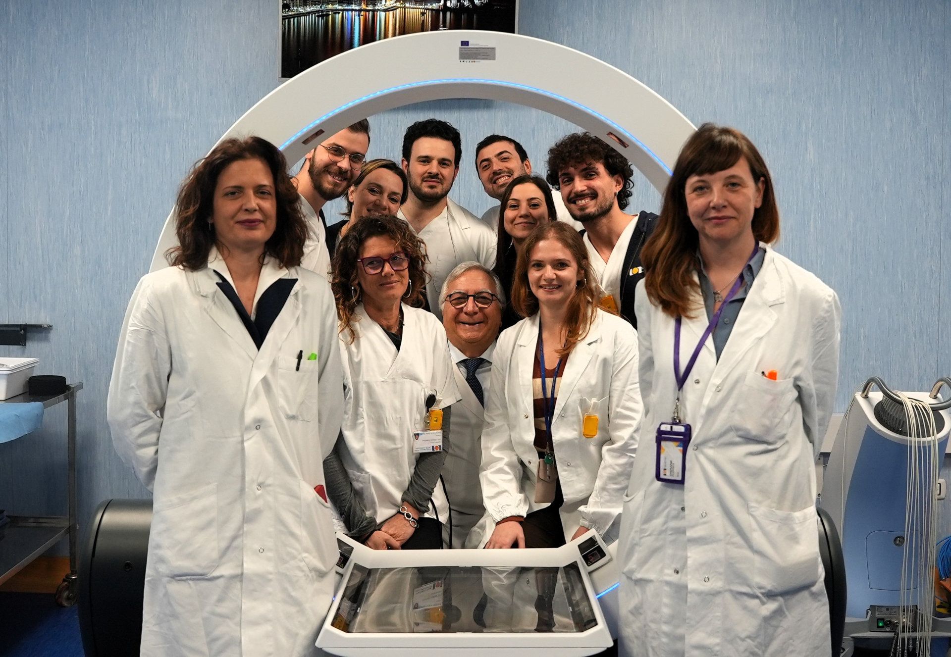
Naples’ NCI Fondazione Pascale interventional radiation oncologists use ImagingRing to complete entire brachytherapy procedure in just one room
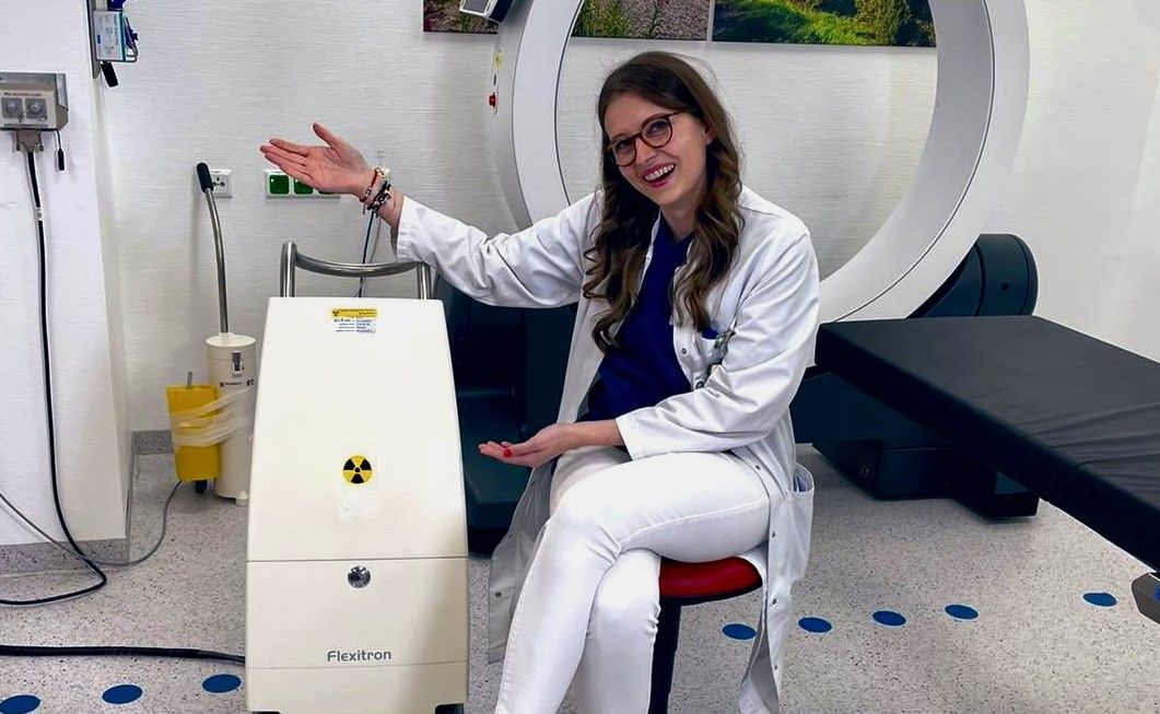
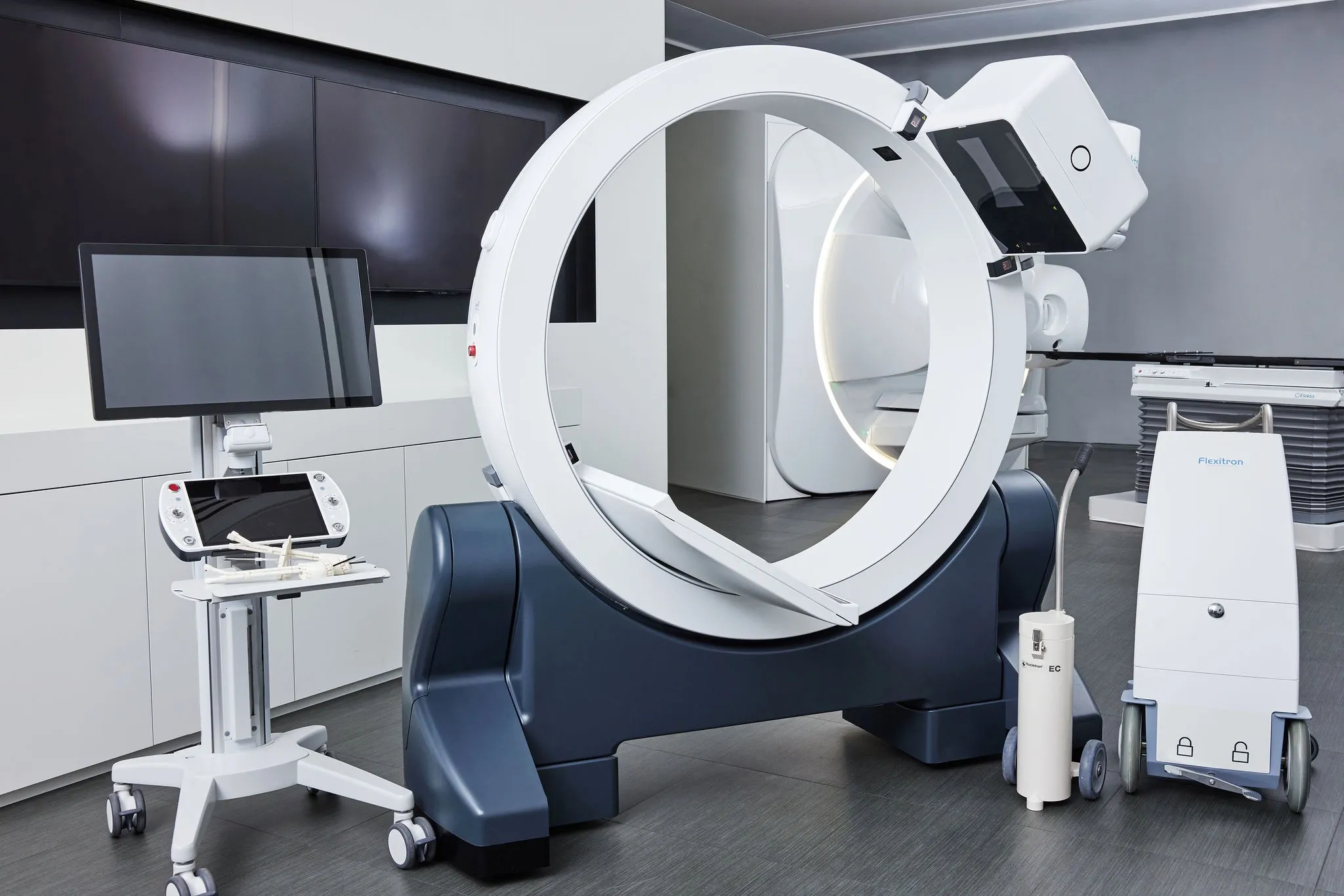
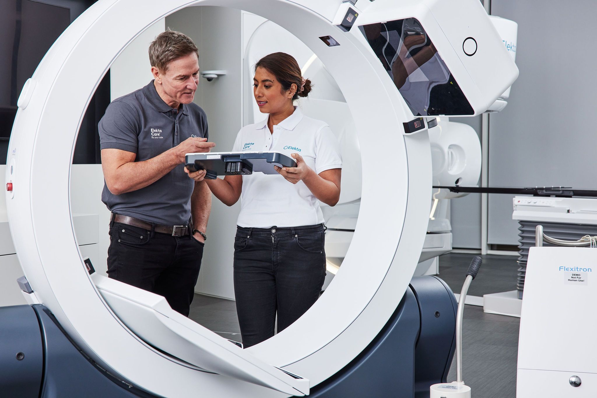
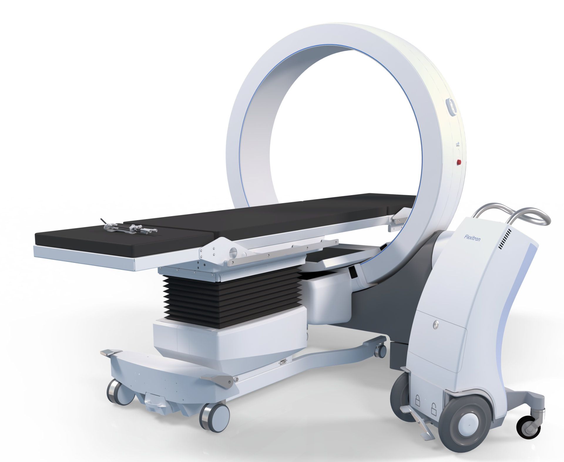
The October 2018 installation of an Elekta Flexitron afterloader facilitated efficient gynecological and skin cancer brachytherapy at NCI Fondazione Pascale (Naples, Italy), and in November 2023 the center experienced an even more dramatic boost in productivity with the acquisition of the ImagingRing. The mobile 3D CBCT imaging component of Elekta Studio, ImagingRing enables NCI Fondazione Pascale interventional radiation oncologists to image, plan, adapt and treat patients in a single dedicated room.
“We acquired ImagingRing for several reasons,” says radiation oncologist Federica Gherardi, MD. “These include improving the accuracy and efficiency of brachytherapy treatments, facilitating the use of interstitial techniques in gynecological implants, shortening both planning and treatment times, and – very importantly – reducing patient discomfort during the brachytherapy process, as the patient can remain in one room for all clinical activities.”
Between October 2018 and 2024, the center’s gynecological and skin cancer brachytherapy volume has steadily increased, reaching a total of 766 patients, comprising 646 gynecological cancer cases (Figure 1) and 120 skin cancer cases (Figure 2). The most significant growth occurred when ImagingRing came into use in late 2023; the annual patient volume leapt from 124 cases to 168 cases.
“ImagingRing also expanded our treatment approach for cervical cancer,” adds Dr. Gherardi’s colleague, radiation oncologist Eva Iannacone, MD. “Before ImagingRing, we focused on post-hysterectomy vaginal cuff and uterine cancer cases using intracavitary brachytherapy only, but in 2024 we were able to include gynecological interstitial implants.”
Single-room brachytherapy workflow better for the patient and the daily schedule
Although brachytherapy with an advanced afterloader is known as a highly effective, focused treatment, the traditional workflow is made more challenging by the need to transport the patient for imaging at various points.
“The traditional workflow for an interventional radiotherapy treatment includes the preparation phase and the implant procedure in the dedicated room, then transport of the patient to the CT room for image acquisition, followed by moving the patient to the treatment room,” Dr. Gherardi says. “However, at Pascale the CT system has been used for all radiotherapy treatments – you can imagine the traffic and the time wasted waiting for a CT slot and the patient’s discomfort having to wait with an applicator inserted.”
The single-room configuration of ImagingRing has eliminated these logistical and patient comfort challenges.
“By removing the need to transport patients for a CT, procedure times have significantly decreased.”
“By removing the need to transport patients for a CT, procedure times have decreased by 40 to 60 minutes, which minimizes any patient discomfort related to the applicator or maintaining the treatment position,” Dr. Iannacone observes. “In addition, in-room CBCT enables treatments under sedation in one room equipped with an anesthesia machine and eliminates concerns about space constraints for the necessary instruments and the brachytherapy team. This is made possible by the compact design of the ImagingRing – which requires just two square meters [21.5 square feet] – allowing for smooth, collaborative work.”
ImagingRing delivers the image quality needed
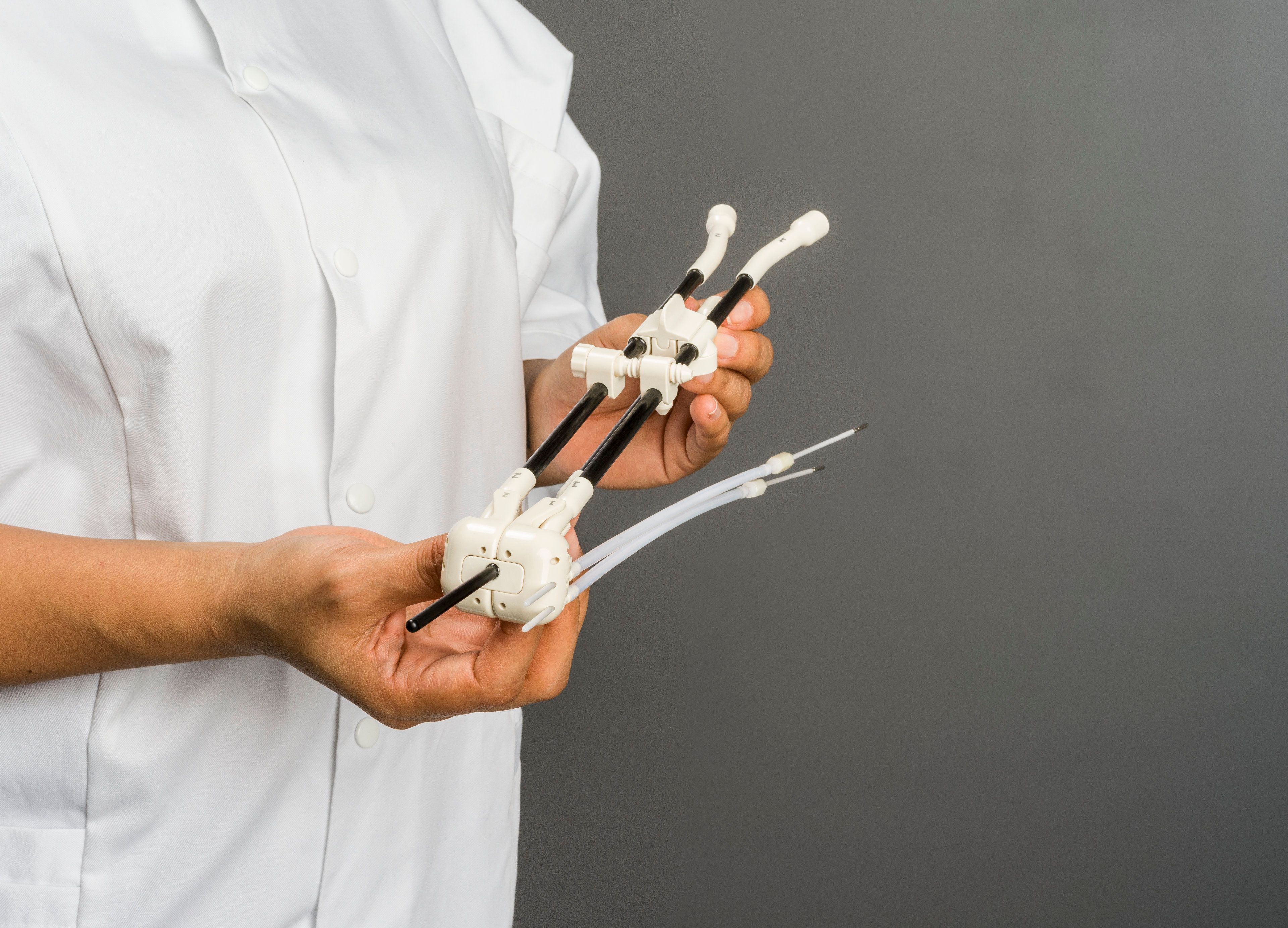
Although a traditional CT provides slightly better image quality and fewer artifacts than the in-room CBCT, the latter – compared to ultrasound – delivers a more precise reconstruction of the brachytherapy applicators and any needles used for interstitial implants, according to the Pascale clinicians.
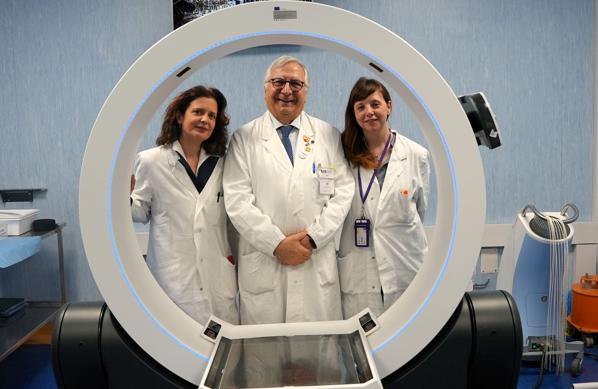
“While we cannot expect superior tumor tissue or OAR definition during the contouring phase compared to MRI, the ability to acquire images directly in the room – quickly and with the patient in certain anatomical positions [e.g., lithotomy position] – offers the advantage of complementing MRI acquisitions,” Dr. Gherardi says.
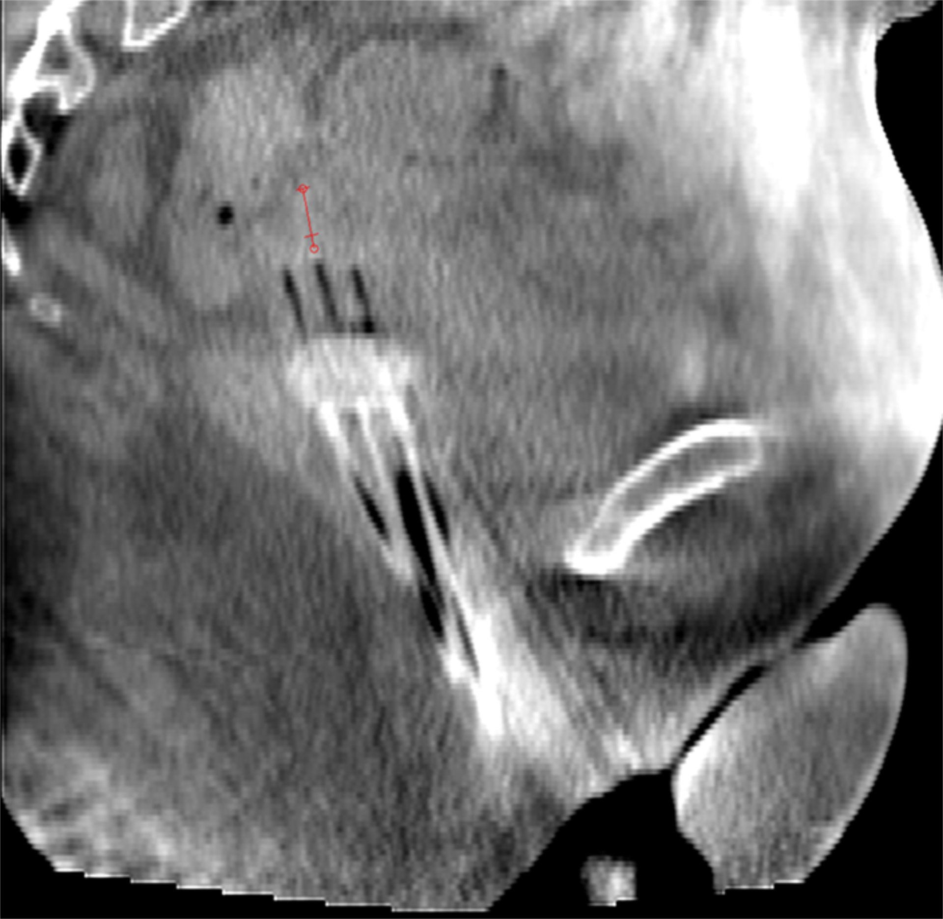
The ability to acquire 3D images helps the brachytherapy team define the target more precisely, enabling improved contouring of both the target and OARs for axial, sagittal and coronal reconstructions, Dr. Iannacone adds.
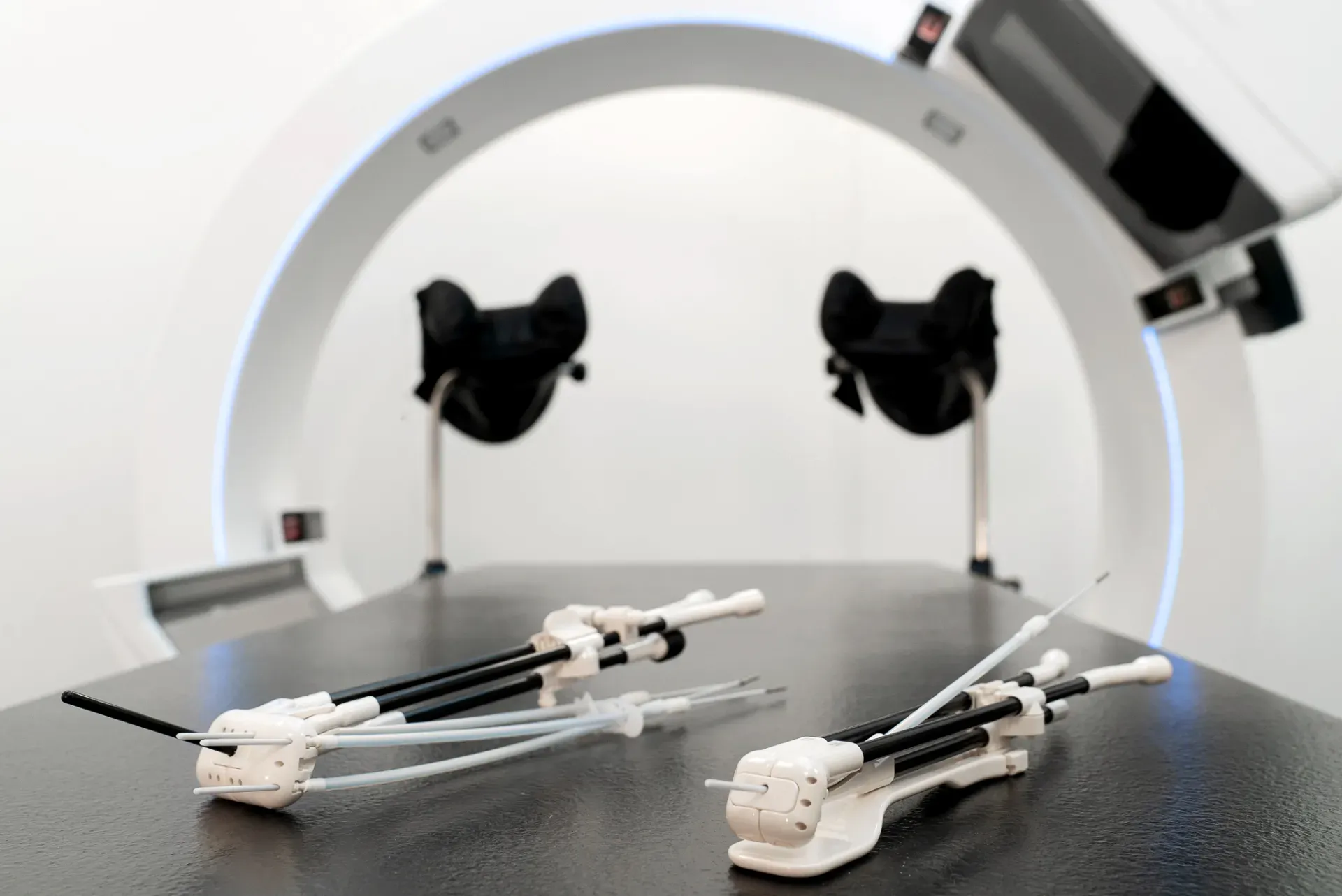
“It also allows us to perform image fusion with diagnostic CT or PET scans, as well as with CBCTs from different treatment fractions,” she explains. “Plus, it facilitates easy reconstruction of applicators, even in more complex implants that involve multiple catheters and/or needle combinations.”
For uterovaginal interstitial treatments, the acquisition of multiple CBCT images throughout the procedure offers guidance for needle placement in the parametrium, enabling Pascale physicians to determine the position and depth of insertion based on the 3D reconstructed images obtained in the implantation position.
“Moreover, in clinical practice – particularly with uterovaginal implants – after the initial procedure, we may find that the applicator isn’t suitable for the patient’s anatomy or extent of the disease,” Dr. Gherardi says. “For example, the applicator’s ovoids may be too small, the endocervical probe may be too short, or the applicator may be poorly anchored.
“…with the ImagingRing we can make immediate adjustments to the implant without the need to move the patient or wait for a CT slot.”
“Before the ImagingRing in these situations,” she adds “it was necessary to return the patient to the interventional room to reposition the applicator and move the patient back to the CT suite for new imaging. However, with the ImagingRing we can make immediate adjustments to the implant without the need to move the patient or wait for a CT slot.”
Dose delivery
Dose delivery occurs about 30 minutes after acquiring the CBCT, which is closely aligned with the planning process. This reassures the Pascale team that the treatment plan remains less dependent on potential intrafraction variations in the OAR positions, on the possible displacement of applicators, or voluntary or involuntary patient movement due to discomfort.
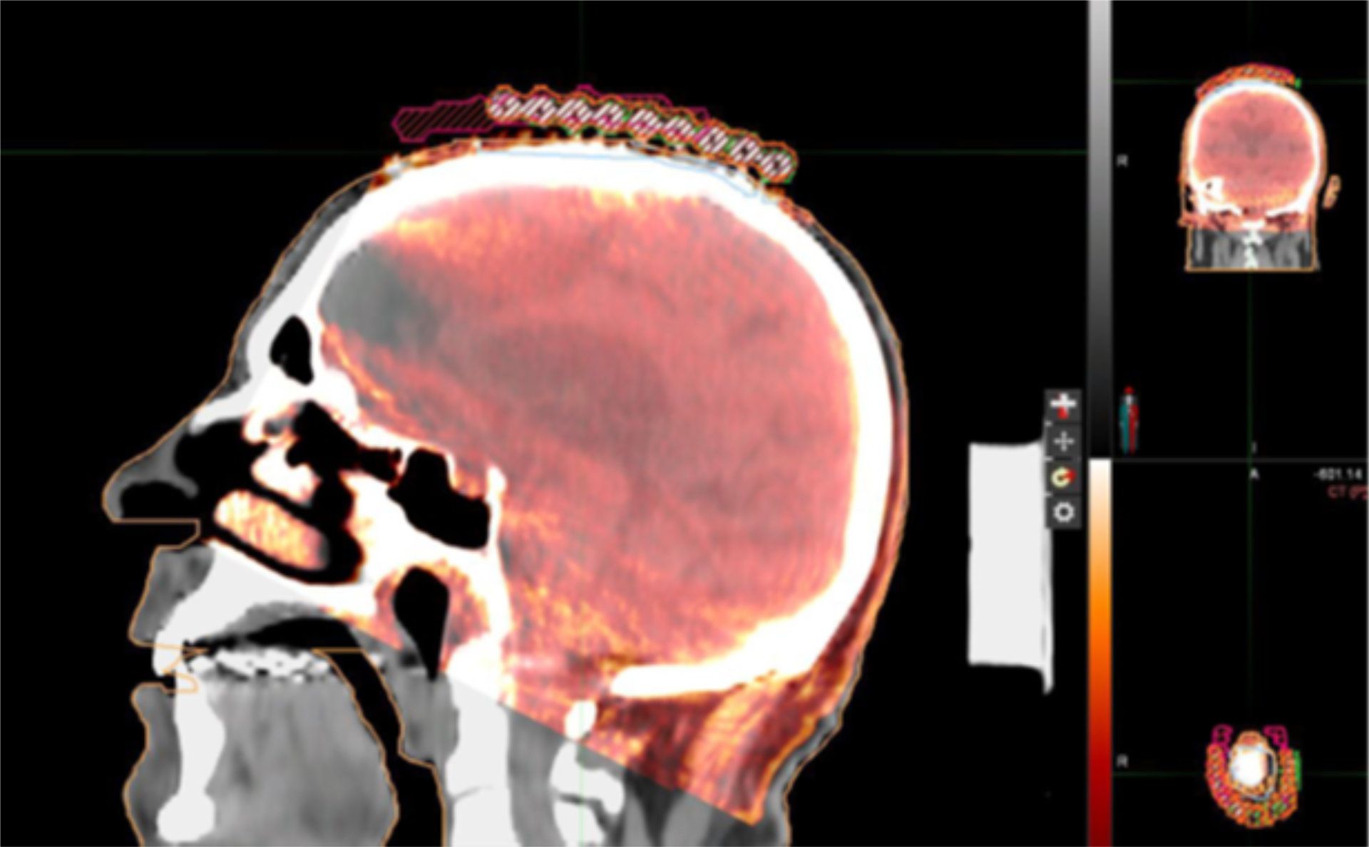
The ability to acquire as many 3D CBCT images as needed has also enabled clinicians to adjust skin treatments if the applicator’s positioning differs from the initial CT, or – in cervical treatments – to modify the plan after changes in rectal or bladder filling, or to adjust the applicator for a more optimal treatment.
“To date – because we are achieving higher quality implants – we have not had to reduce the prescribed treatment dose,” Dr. Gherardi notes.
Post-procedure CBCT confirms plan suitability
Immediately after dose delivery, Pascale radiation oncologists routinely perform a repeat CBCT to monitor intra-fraction and applicator motion to assess the adequacy of the plan at the end of each fraction.
“This reassures us about the feasibility of re-delivering the first fraction in simpler cases, such as the treatment of the vaginal cuff,” Dr. Iannacone says. “By evaluating the applicator and surrounding OARs via a scout image before dose delivery, we can now proceed with image fusion with the first fraction plan CBCT and perform instant replanning only when necessary.”
Patient comfort a priority
While the ImagingRing makes the lives of clinicians much less hectic by anchoring them to one room, Pascale clinicians focus mainly on how a single-room procedure positively impacts the patient experience.
“Above everything else, patient comfort represents the true advantage of the ImagingRing.”
“Above everything else, patient comfort represents the true advantage of the ImagingRing,” Dr. Gherardi asserts. “It offers the possibility to simplify the workflow by eliminating all phases of patient mobilization, as well as performing the entire procedure under sedation, so that the patient has no perception of any part of the process.
“The patient is sedated while lying down before the insertion of the catheter,” she continues, “and is awakened once the treatment is delivered and the applicator and catheter are removed. The patient then attends subsequent interventional radiotherapy sessions without anxiety and no memory of discomfort.”
Looking toward the future
In addition to incorporating MRI acquisitions – fusing them with CBCT images using Oncentra Brachy image fusion software to better define the target – Pascale clinicians are also exploring how the ImagingRing could be employed for new indications.
“We would like to implement interstitial breast treatments with the collaboration of interventional radiologists for endoluminal interventional radiotherapy, such as bronchial or biliary applications,” Dr. Iannacone says. “With the availability of in-room imaging, procedure times and the duration of anesthesia can be reduced. Furthermore, catheter positioning could be fluoroscopically guided and checked before acquiring and transferring CBCT images to the planning system.”
Dr. Gherardi adds that the ImagingRing offers substantial value for centers where optimizing the efficiency of the entire radiotherapy workflow is essential.
“We highly recommend the ImagingRing for clinics with high patient volumes.”
“We highly recommend the ImagingRing for clinics with high patient volumes,” she says. “And also for centers trying to reduce patient anxiety and discomfort associated with interventional procedures, particularly for patients undergoing invasive treatments and requiring sedation.
“The ImagingRing enables these procedures to be performed without the patient experiencing anticipatory distress or pain,” Dr. Gherardi adds, “thus providing a smoother, more comfortable experience for patients and staff. Finally, we recommend ImagingRing for hospitals with multiple treatment rooms within the same department, as it allows for time optimization by enabling the system to be easily moved between rooms.”
Click here to learn more about image guided brachytherapy with Elekta Studio Imaging Ring.
Elekta is the authorized Exclusive Distributor of medPhoton in brachytherapy. ImagingRing is product manufactured by medPhoton GmbH. ImagingRing may not be available in all markets.
LARBX250506

