Enhanced confidence for prostate SBRT with Clarity Autoscan
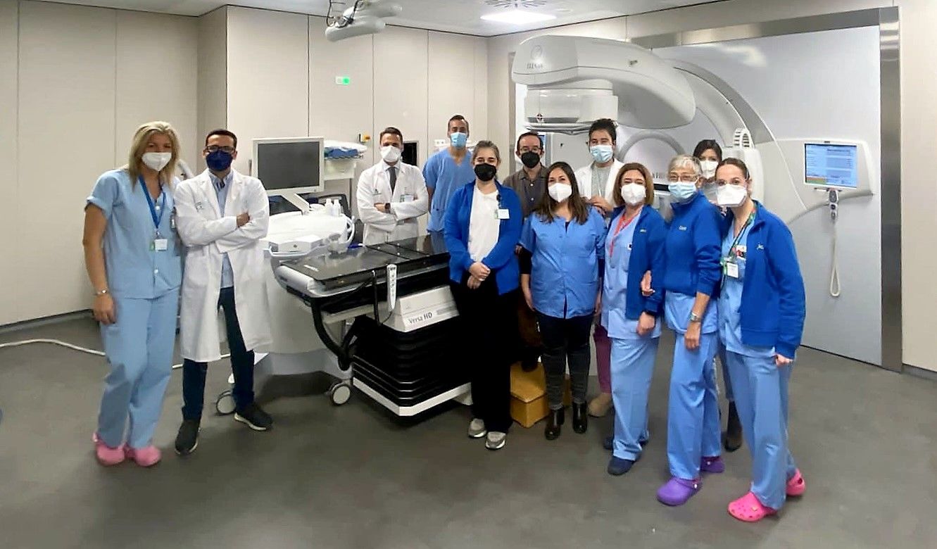
The University Hospital Virgen Macarena in Seville, Spain, reduces margins for prostate SBRT thanks to real-time ultrasound imaging with Clarity Autoscan
In October 2019, the radiation therapy department at the University Hospital Virgen Macarena (UHVM) in Seville installed Clarity® Autoscan with a view to implement prostate SBRT. By introducing real-time 4D ultrasound monitoring of prostate movement during treatment delivery, they hoped to benefit-eligible prostate SBRT patients by increasing treatment accuracy, shrinking treatment margins and reducing irradiation of healthy tissues and organs at risk (OARs).

Previously, the department employed a moderate hypofractionation schedule (60 Gy in 20 fractions) to treat prostate cancer patients, using intraprostatic fiducials for prostate localization. CBCT images were taken daily for patient setup for the first few fractions and then weekly for the remaining fractions, or more frequently depending on the mobility of the prostate. Seeing Clarity in use at the University Hospital HM Sanchinarro in Madrid gave the UHVM team confidence that real-time intrafraction ultrasound imaging of the prostate gland would help them ensure accurate dose delivery for prostate SBRT.
“We are used to visualizing the prostate in CT scans, but transperineal ultrasound imaging is quite different,” explains Dr. Maria Rubio, Radiation Oncologist at UHVM. “Seeing Clarity in the hands of an experienced team at the HM Sanchinarro Hospital was extremely helpful. In return, Elekta experts came to our department to train staff on the equipment and help them get used to the ultrasound images. After one week, we began to use Clarity clinically.”
Real-time monitoring
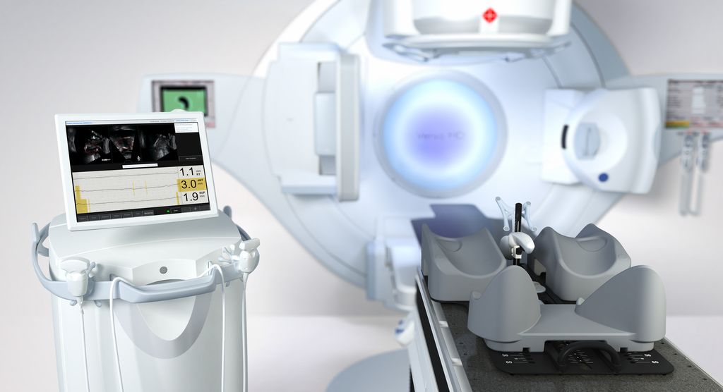
For the implementation of the prostate SBRT program, staff were fully trained on Clarity and followed the department’s prostate SBRT protocol, which is based on NCCN guidelines. Clarity 4D monitoring provides live imaging of the prostate and surrounding anatomy during treatment. The system automatically calculates target position compared to physician-defined thresholds, enabling precise intrafraction motion management.
“Clarity is used for real-time intrafraction monitoring for all five fractions, giving us confidence to deliver high doses accurately.”
“For prostate SBRT, we deliver 36,25 Gy in five fractions,” says Dr. Rubio. “Clarity is used for real-time intrafraction monitoring for all five fractions, giving us the confidence to deliver high doses accurately.”
“In the simulation phase, we perform a CT scan and acquire transperineal ultrasound images using Clarity to localize the prostate and fuse with the planning CT scan,” she continues. “On the first day of treatment, patient setup is performed using CBCT. Then we monitor prostate movement in real-time during beam delivery using Clarity. With automatic beam gating, if the prostate moves outside of our pre-defined limits for more than five seconds, the beam stops. The position of the prostate is corrected, and alignment confirmed using Clarity before treatment delivery is resumed.”

Protecting organs at risk
The precise intrafraction motion management provided by Clarity has enabled the team to reduce their treatment margins.
“With our 20-fraction hypofractionated schedule, we were using treatment margins of 8-10 mm,” Dr. Rubio comments. “Now, using Clarity for SBRT, our margins are 3-5 mm. We know that with real-time monitoring if the prostate moves outside of our tolerances, the beam will stop. This intrafraction control of the beam gives us the assurance we need to deliver SBRT doses safely and accurately. We can be sure that the prostate position is within the limits we have set during each treatment fraction.”
“This intrafraction control of the beam gives us the assurance we need to deliver SBRT doses safely and accurately.”
The use of tighter treatment margins lowers the risk to patients by reducing dose to surrounding healthy tissue and OARs.
“We have found that the side effect profile for prostate SBRT is no different to our previous moderate hypofractionated schedule, despite the use of higher doses per fraction,” Dr. Rubio adds. “Also, since some of our patients have some distance to travel, they are very happy to have their treatment completed in fewer sessions.
“Once you start using Clarity for prostate SBRT, you will never want to stop.”
“Once you start using Clarity for prostate SBRT, you will never want to stop,” she concludes. “It has allowed us to ensure treatment accuracy throughout each application, providing a safe and convenient treatment option for our patients.”
Learn more about how Clarity can support you and the prostate SBRT program in your clinic.
Real-time soft tissue tracking with Clarity® Autoscan
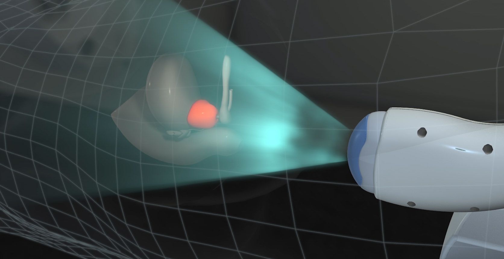
Elekta Clarity® Autoscan provides live 4D monitoring of the prostate and surrounding anatomy during treatment, without the need for surgical implants or ionizing radiation. Using real-time ultrasound imaging, prostate position is automatically calculated and compared to physician action thresholds, enabling precise intra-fraction motion management and accurate dose delivery.
LWBCLA220225
