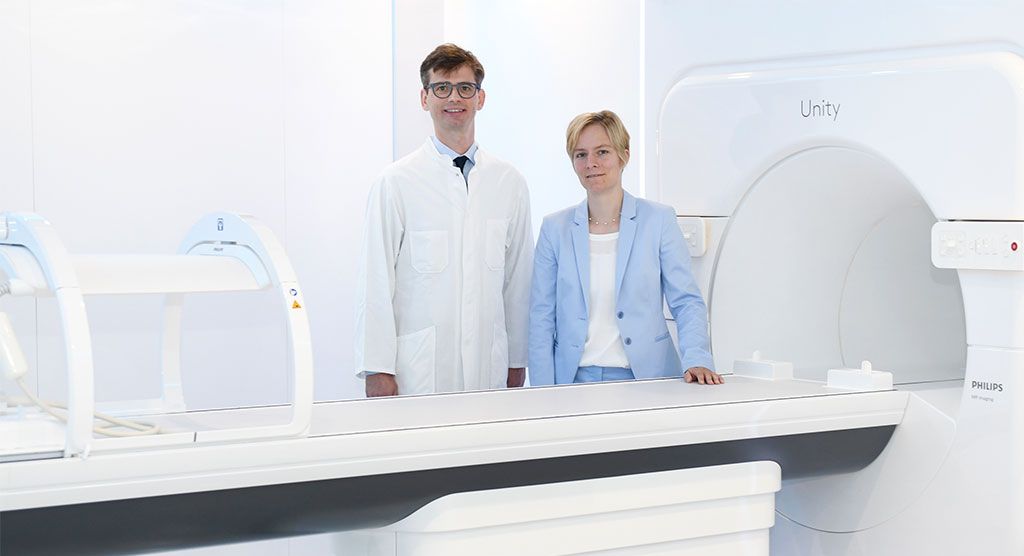
Elekta Unity reveals advantages of real-time imaging during radiotherapy
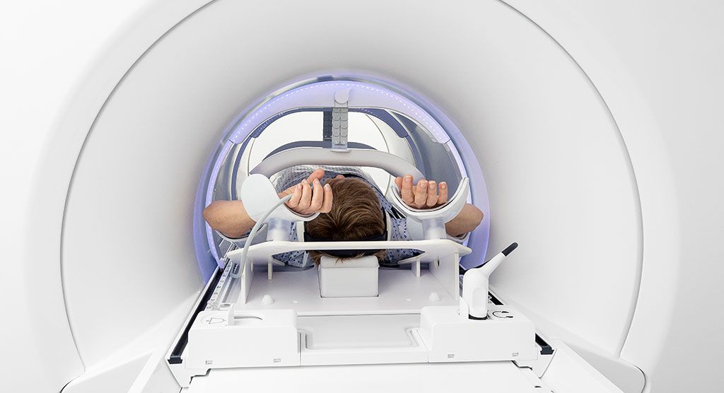
Marking a year of use, presenters in Elekta-sponsored webinar review impact of 3D MRI-guided adaptive radiotherapy
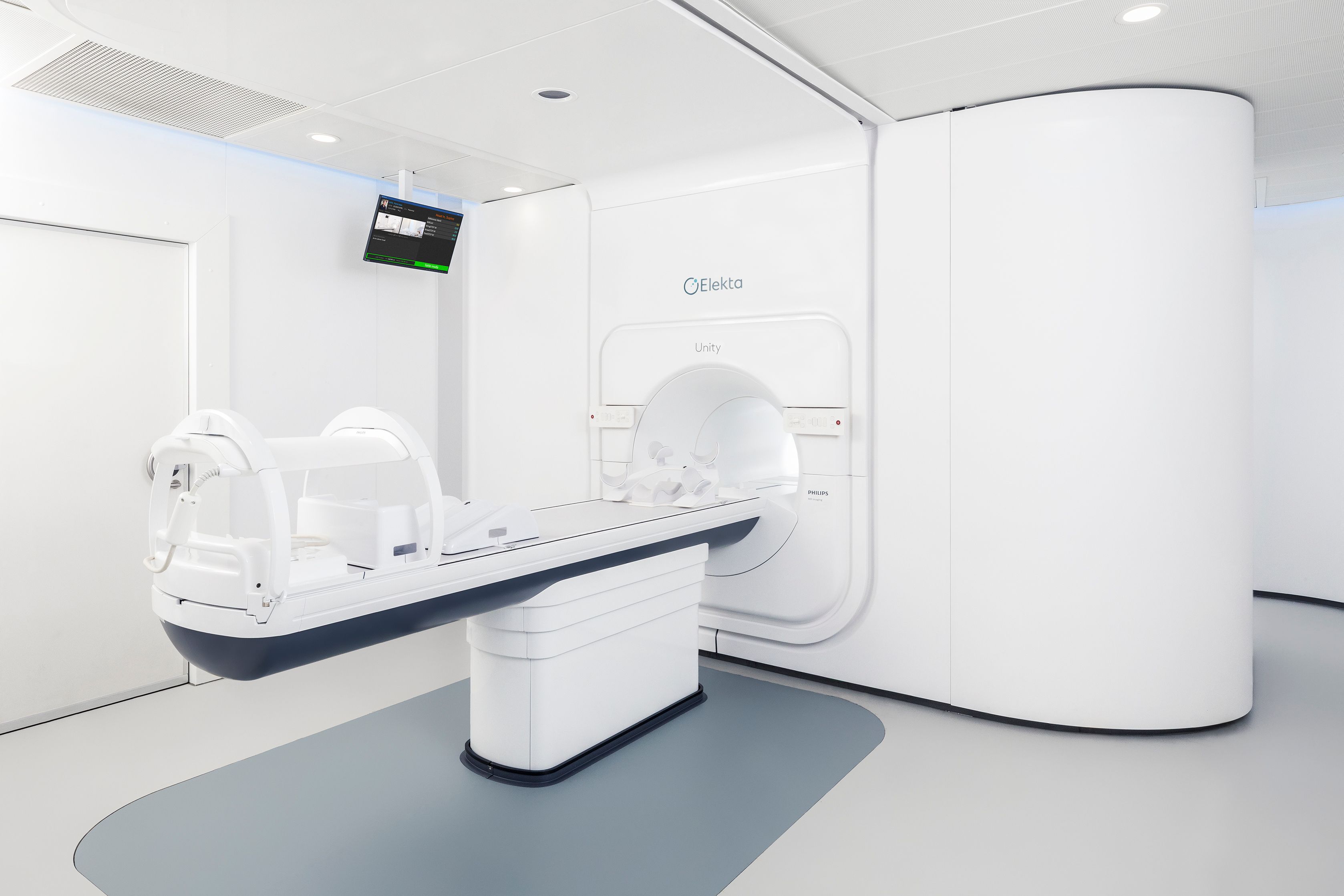
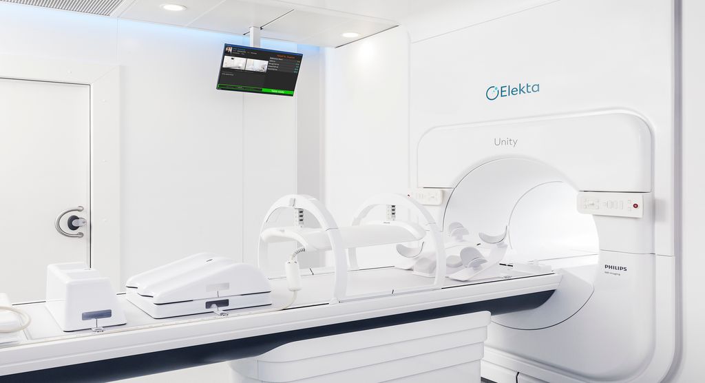
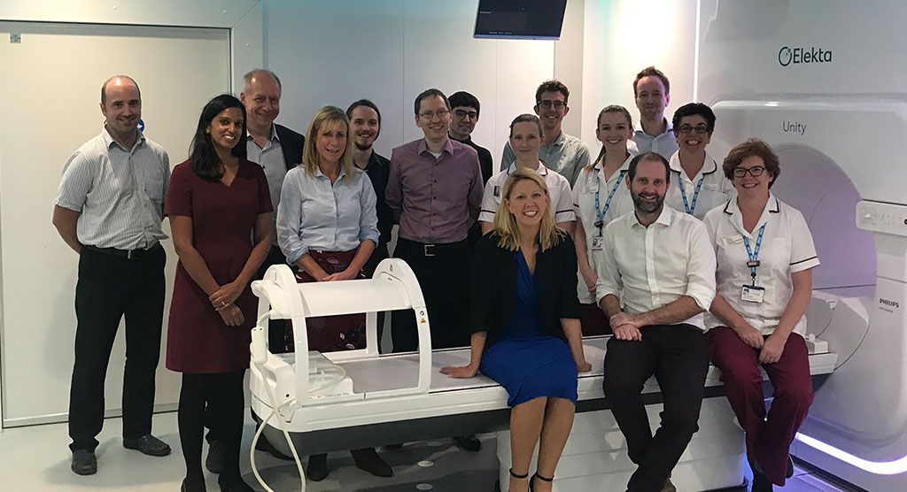
The ability to see both tumor targets and surrounding healthy tissue with high quality MR imaging while delivering radiation is having a profound impact at oncology departments worldwide. During a webinar hosted by Physics World, Alison Tree, MD, at the Royal Marsden (UK) and Cihan Gani, MD, at the University Hospital Tübingen (Germany), discussed how Elekta Unity MR-Linac is changing the practice of radiotherapy. View their presentation, titled “Celebrating one year of clinical activity with Elekta Unity.”
Dr. Tree, consultant clinical oncologist at the Royal Marsden, observed that traditional imaging in RT planning has been less optimal in most cases, the depiction of targets and OARs being “a bit like staring at a boat across a foggy harbor.”
In prostate imaging, for example, kV imaging can accurately visualize prostate fiducials, but provides no information about bladder or bowel filling relevant to side effects and toxicity. With the MRI technology in Elekta Unity, however, she can clearly see prostate borders and capsule, the key to reducing margins.
“The same applies to the female pelvis,” Dr. Tree added. “Using CBCT, it’s very difficult to distinguish different tissues, where with MRI you can easily distinguish the bladder, uterus and rectum.”
Using MRI to clearly visualize tumor targets and healthy tissues during the planning process is an obvious advantage, Elekta Unity also provides that same MRI image quality in real-time during treatment. That makes the system a game-changer.
“With Elekta Unity, I can visualize the prostate-rectal interface well enough during beam delivery that I can see if the [prostate] is moving out of the way of the radiation.”
“In conventional radiotherapy, at the very moment that we most need precision, we turn all our imaging off,” she said. “With Elekta Unity, I can visualize the prostate-rectal interface well enough during beam delivery that I can see if the [prostate] is moving out of the way of the radiation.”
Dr. Tree reviewed several male and female pelvic malignancy cases in which bladder/bowel filling caused target movement or deformation. She added that tumor shrinkage during the radiotherapy course also must be taken into consideration.
“What the MR-Linac allows us to do is change our plan on a daily basis to account for any anatomical changes,” she said.
Throughout the Royal Marsden’s first year of Elekta Unity activity, Dr. Tree recorded some relevant statistics:
MR-Linac treatment time
| Tumor site | Average (SD) time on treatment couch (mins) |
|---|---|
| Prostate | 39:13 (3:09) |
| Rectum | 36:25 (1:31) |
| Bladder | 41:23 (1:53) |
| Gynecological | 23:50 (2:26) |
Timings breakdown per activity in workflow (prostate)
| Workflow step | Time (min.) |
|---|---|
| Patients set up | 4.0 |
| Pre-treatment scan and registration | 6.0 |
| Contouring | 8.5 |
| Preparing for optimization | 3.5 |
| Optimization | 4.0 |
| QA and plan sign (now concurrent) | 6.0 |
| Beam on | 4.0 |
| Post-treatment imaging | 4.0 |
The University Hospital Tübingen’s Dr. Gani reported that by September 2019 his center had used Elekta Unity to treat over 80 patients with a total of more than 1,200 fractions. Apart from prostate and rectal cancers, head-and-neck tumors and partial breast irradiation, liver malignancies have been the main indication.
Elekta Unity is CE-marked and 510(k) cleared. Not commercially available in all markets.
DWI is CE-marked and pending FDA 510(k) for brain applications. Intended for imaging purposes.
Diagnostic image quality and imaging during delivery are pending 510(k) with FDA.
