Clarity Autoscan improves radiotherapy delivery accuracy for prostate cancer
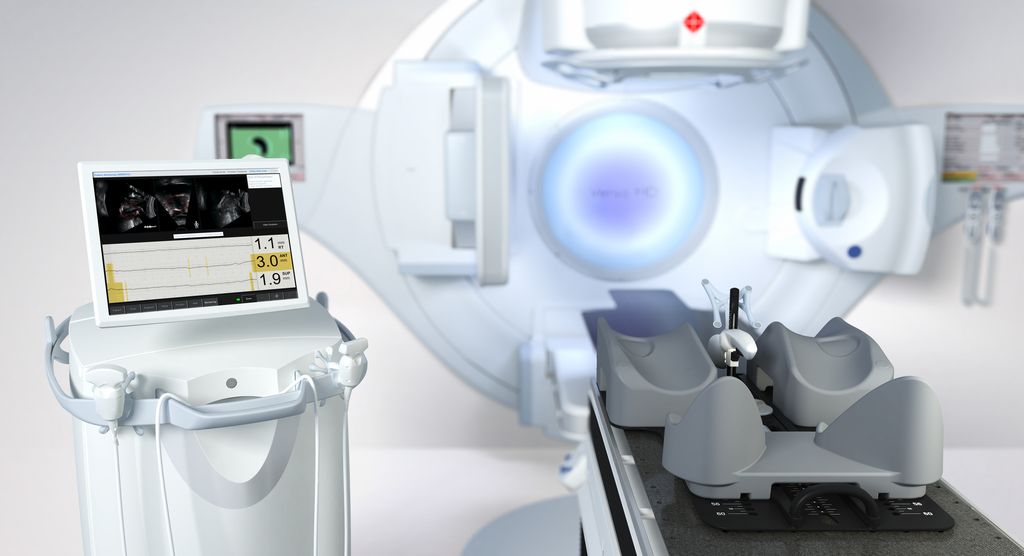
The First Affiliated Hospital of Xi’an Jiaotong University in China is first in country’s northwest region to debut ultrasound-guided technology
Due largely to China’s aging population, prostate cancer has emerged as the sixth most common cancer in the country1, driving a surge in prostate cancer patients at the nation’s medical centers. To enable more accurate radiotherapy treatments of individuals with prostate cancer, The First Affiliated Hospital of Xi’an Jiaotong University began using Elekta’s Clarity® Autoscan soft-tissue visualization system in November 2018. The solution enables clinicians to monitor prostate position in real time by positioning an ultrasound probe against the patient’s perineum.
“The ability to track prostate position and movements in real time and with submillimeter accuracy is critical.”
“The ability to track prostate position and movements in real time and with submillimeter accuracy is critical, because the prostate can move up to 2.5 cm during treatment, due to rectal and/or bladder filling,” says Zhang Xiaozhi, MD, Director, Radiotherapy Department, First Affiliated Hospital of Xi’an Jiaotong University.
“If we can monitor prostate position before and during radiotherapy delivery, we can ensure that the radiation is correctly delivered to the target tissue and not to sensitive, healthy tissues nearby, such as the rectum and bladder. The other major benefit is that using ultrasound is noninvasive and doesn’t use ionizing radiation.”
He adds that because Clarity Autoscan improves radiotherapy delivery accuracy, it will be possible to increase the dose per treatment session.
“If we can deliver more potent therapeutic doses for each fraction, we should be able to reduce the number of treatment sessions the patient requires,” Dr. Xiaozhi says. “Clarity Autoscan represents a major new improvement in our ability to treat this increasing patient population.”
Case study: 66-year-old patient with prostate cancer
For this patient’s case, clinicians had not initially considered intra-fraction imaging and motion management for his treatment. However, after researching the literature, they realized that Clarity Autoscan would enable intra-fraction imaging and real-time monitoring during beam delivery.
“After discussing the patient’s case with family members, we opted to use Clarity Autoscan to treat the patient,” Dr. Xiaozhi says.
For ultrasound localization, the patient is comfortably immobilized in the treatment position, and the prostate ultrasound probe is gently placed against the patient’s perineum to provide a clear viewing window to the target region. The physician is free to place the probe in the best position to view the prostate, allowing clear visualization of the prostate’s position. Then the ultrasound positioning scan is performed.
Following localization, a CT scan is acquired. The CT and ultrasound images are transferred to the ultrasound guidance system and the images are automatically fused, with Director of Physics Li Yi verifying the fusion. Then, physician, Qu Yiping, contours the prostate target using the fused images. After contouring, the target area and fused CT image are transferred to the treatment planning system.
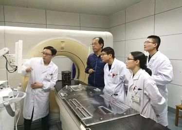
“Compared with CT positioning image only, the fused image with Clarity Autoscan is much clearer for us to contour the target and we can reduce the margin to better protect the critical structures,” Li Yi, Director of Physics, confirms.
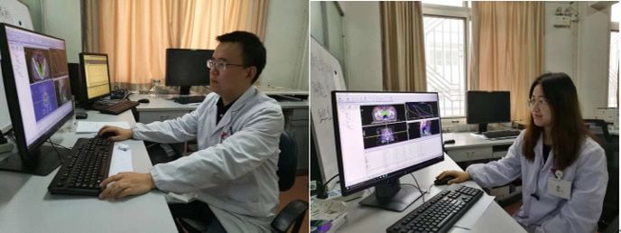
The clinician uses the fused image to contour the tumor target and normal tissues. Physicists Zhang Yuemei and Yi employ Monaco® treatment planning system to create several VMAT treatment plans, and then consult with the physician to select the best therapeutic regimen.
Before treatment, technicians Wang Long and Zhang Jiadi conduct ultrasound system daily quality testing, confirming that the system can achieve submillimeter accuracy.
Therapists reposition the patients based on the treatment plan, achieving alignment between treatment isocenter and tumor isocenter. Then, cone beam CT positioning is done, and Clarity monitors and maintains the planned position. Finally, the HexaPOD™ evo RT System’s robotic couch is automatically adjusted based on the setup errors and corrects the patient’s position for treatment.
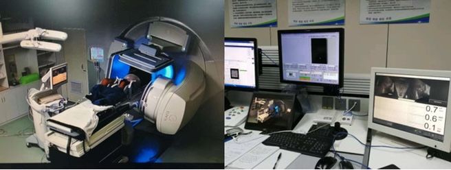
During treatment, the clinicians can observe the real-time scanning images in the control room, with Clarity pausing the beam automatically when predefined thresholds are exceeded. The treatment will be resumed based on the real-time prostate positions detected by Clarity, making sure the target area is always within the treatment beam.
The First Affiliated Hospital of Xi’an Jiaotong University introduced Clarity Autoscan, which provides high-quality and clear imaging of the prostate and surrounding soft tissues, real-time tracking of the motion during treatment and helps protect critical structures. The implementation of this new technology will bring noninvasive treatment to more prostate cancer patients.
“Elekta Clarity Autoscan is safe and reliable,” Dr. Xiaozhi says. “We can see so much with it, and make corrections through real-time tracking, to make sure doses are delivered to the target accurately and critical structures are well protected.”
1National Cancer Incident Registration Annual Report 2017
