A growing global challenge
Brain tumors affect more than 320,000 people each year, with nearly 250,000 deaths worldwide. For many patients, radiosurgery and radiotherapy are the most effective and least invasive options. Adaptive treatments offer a personalized approach, bringing safer, more effective care to more patients.1
people affected each year
Proven precision, smarter protection
Key clinical evidence shows that adaptive radiosurgery and radiotherapy enables accurate, personalized treatment for brain tumors, maximizing target coverage while protecting critical structures.
3 cm tumor change
Glioblastomas can shrink or shift by as much as 3 cm over the course of treatment due to edema, tumour regression, or edema, underscoring the need for adaptive radiotherapy to keep pace with these changes.2
Tumor progression during delays
A two-week gap between imaging and treatment led to new brain metastases in 50% of patients and tumor growth in 75% with longer delays linked to greater increases.3
Improved sparing of motor, sensory and neurocognitive function
Radiosurgery for multiple brain metastases can support stabilize and even improve cognitive function.4
How adaptive radiotherapy helps you respond to clinical changes
| Your challenge | Adaptive helps you... |
| Anatomy changes during the treatment course | Re-optimize daily treatment to stay on target and reduce unwanted dose to healthy tissue |
| Tumors shrink or move between treatment sessions | Adjust treatment plan based on the image at the time of treatment |
| Escalating dose without treating organs at risk | Achieve greater certainty of patient position and adapt to what you can see on the day |
| Quick replanning for scan-plan-treat workflows | Improves patient throughput and reduces time to treatment so more patients treated promptly |
Clinics are already adapting and redefining what’s possible
Nobody does adaptive like Elekta
Treating brain tumors and other lesions is always complex and we're here to support you. We don't just enable advanced adaptive radiotherapy and stereotactic radiosurgery, Elekta enables sub-millimetre precision and daily adaptation to anatomical change. Beyond technology, we partner with clinicians worldwide to help preserve cognitive and neurological function while delivering the precision every patient deserves.

The brain demands extraordinary precision
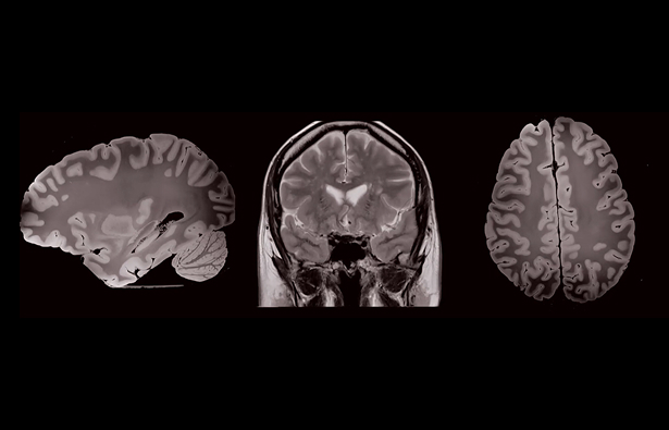
Tumors in the brain like glioblastoma, brain metastases, meningiomas or acoustic neuromas, can change from session to session. Even subtle changes in anatomy can affect how much healthy brain tissue is included within the treated area.
As the whole brain is a critical structure it is crucial to keep dose outside the target to a minimum, protecting cognitive motor and sensory movement, you can’t afford to compromise.
Adaptive radiosurgery helps you stay on target. With pre-treatment imaging and dose reoptimization, you can adapt to what’s changed, reduce, or even eliminate margins, and deliver treatment with confidence.5
Adaptation gives you more options, more control
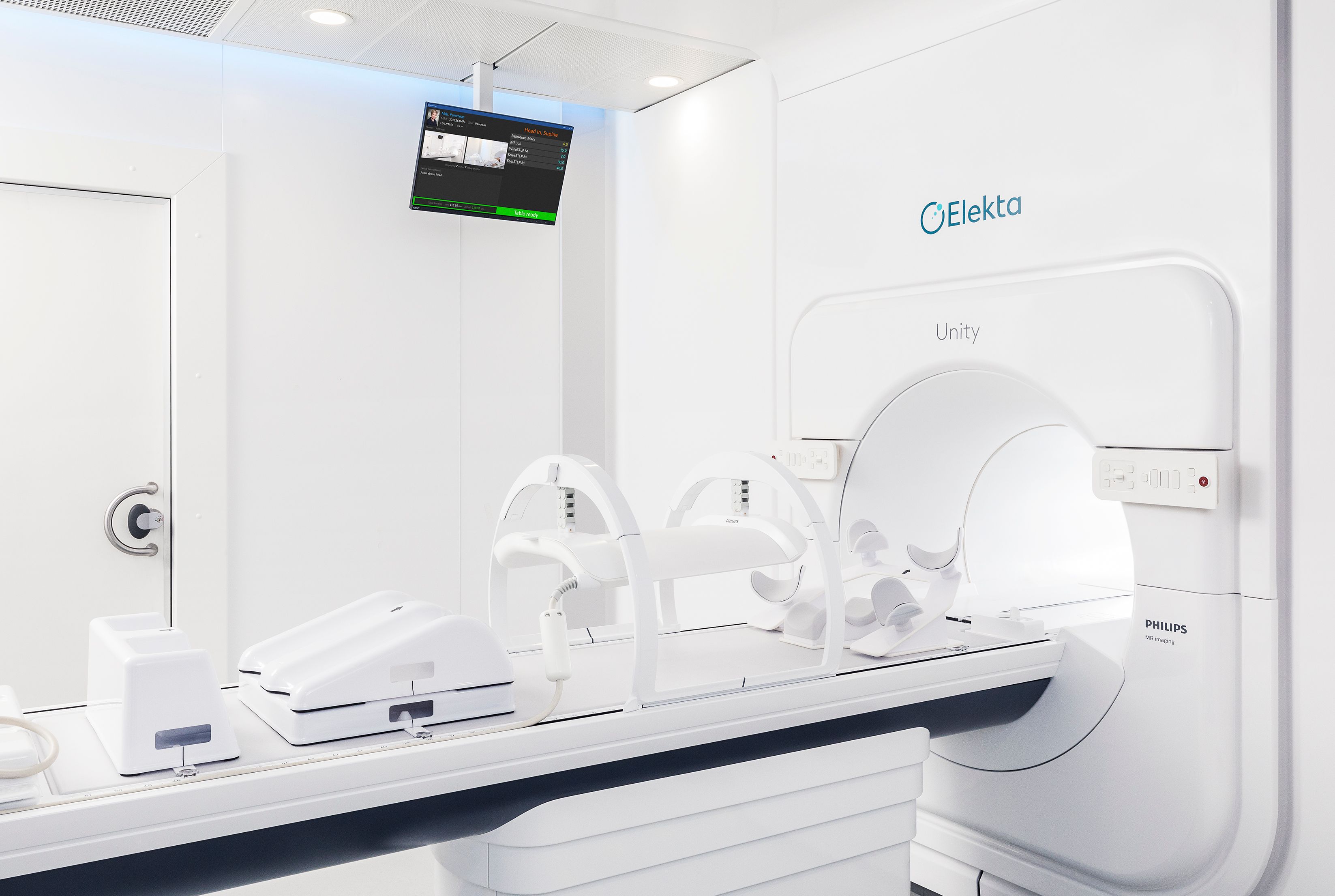
Daily adaptive workflows empower your team to respond to what’s changing, whether that’s tumor shrinkage, edema reduction, or anatomical shifts. With clear MR or CBCT images and on-the-fly plan adaptation, you can:
Reduce or eliminate margins
Confidently reduce margins to protect healthy brain tissue, preserving motor, sensory and neurocognitive function.6, 7
Adapt to changing anatomy
Adjust plans to match what you see, helping maintain full target coverage, and reduce dose to organs at risk, as the tumor responds to treatment.8
Simplify workflow
Make plan adjustments faster and more precisely, without the delays and resource burden of offline replanning.
Share your thoughts
We're asking how centers are approaching adaptive radiotherapy, where are you on the journey?
1. Why are you interested in content on adaptive radiotherapy?
Delivering the future of brain tumor treatment
Whether you’re treating glioblastoma, brain metastases, meningiomas, acoustic neuromas, or other benign tumors in adult or pediatric cases, your patient deserves the most accurate treatment possible to ensure long-term quality of life.
With MR and CT options for daily image guidance, and streamlined workflow tools to create efficiency, you can deliver more personalized, precise treatment for your patient, while maintaining high patient throughput and protecting what matters most.
Explore our adaptive solutions for brain tumors
Our image-guided adaptive solutions help clinicians treat with sub-millimeter precision, safeguarding healthy brain tissue and critical structures while maintaining tumor control.
References
- 2025 Global Impact Report: Precision targeting, global impact – cancer radiotherapy in the 21st century. (2025). Available at: https://aboutadaptive.com
(Accessed: 16 October 2025). - Stewart, J. et al. (2021) ‘Quantitating interfraction target dynamics during concurrent chemoradiation for glioblastoma: a prospective serial imaging study’, International Journal of Radiation Oncology, Biology, Physics, 109(3), pp. 736–746. Available at: https://pubmed.ncbi.nlm.nih.gov/33068687/(Accessed: 16 October 2025).
- Cahill J. et al. (2024) ‘Progress of intracranial metastases during the interval before stereotactic radiosurgery: a retrospective cohort analysis’, European Journal of Surgical Oncology, 50(12), 108676. Available at: https://doi.org/10.1016/j.ejso.2024.108676
(Accessed: 16 October 2025). - Schimmel W.C.M., Verhaak E., Bakker M. et al. (2021) ‘Group and individual change in cognitive functioning in patients with one to ten brain metastases following Gamma Knife radiosurgery’, Clinical Oncology (Royal College of Radiologists), 33(5), pp. 314–321. Available at: https://doi.org/10.1016/j.clon.2021.01.003
(Accessed: 16 October 2025). - Gamma Knife Radiosurgery for Brain Metastases - Clinical Leaflet
- Chang E.L., Wefel J.S., Hess K.R. et al. (2009) ‘Neurocognition in patients with brain metastases treated with radiosurgery or radiosurgery plus whole-brain irradiation: a randomized controlled trial’, The Lancet Oncology, 10(11), pp. 1037–1044. Available at: https://doi.org/10.1016/S1470-2045(09)70263-3
(Accessed: 16 October 2025). - Niranjan, A., Monaco, E., Flickinger, J. et al. (2019) ‘Guidelines for multiple brain metastases radiosurgery’, Progress in Neurological Surgery, 34, pp. 100–109. Available at: https://pubmed.ncbi.nlm.nih.gov/31096242/
(Accessed: 16 October 2025). - Tseng C.L., Chen H., Stewart J., Lau A.Z., Chan R.W., Lawrence L.S.P., Myrehaug S., Soliman H., Detsky J., Lim-Fat M.J., Lipsman N., Das S., Heyn C., Maralani P.J., Binda S., Perry J., Keller B., Stanisz G.J., Ruschin M. and Sahgal A. (2022) ‘High-grade glioma radiation therapy on a high-field 1.5 Tesla MR-Linac – workflow and initial experience with daily adapt-to-position (ATP) MR guidance: a first report’, Frontiers in Oncology, 12, 1060098. Available at: https://doi.org/10.3389/fonc.2022.1060098
(Accessed: 16 October 2025).


